Vestibular Testing at UCSF Balance and Falls Center
Videonystagmography (VNG)
VNG is a technique designed to evaluate how the inner ear is functioning. Since one of the main functions of the balance system is to keep our visual world steady during head movements, the eyes and the inner ears are connected through a reflex arc known as the vestibulo-ocular reflex. Therefore, every time the inner ear senses a head movement, the eyes move in an equal and opposite direction. Because of this feature, we are able to use eye movements as a window to study the inner ear. Eye movements can be measured in a variety of ways, including by using electrodes to measure electrical changes associated with movement (ENG, or electronystagmography), or with infrared goggles and computer software that tracks the position of the pupil (VNG). The test is non-invasive, and involves several parts. First, the patient is asked to visually track targets that will either jump around (saccades), or move continuously (smooth pursuit). This part tests the oculomotor system, which is the neural network that controls eye movements. Second, positional nystagmus is assessed, with placing the patient into certain positions that may cause dizziness. This is important to see if the dizziness is related to BPPV. Finally, caloric testing is performed, which involves placing hot and cold air or water into each ear to stimulate the inner ear (mostly the horizontal semicircular canals). The speed of the resulting eye movements (specifically the slow phase of the nystagmus) is then measured. This test is helpful for determining if the inner ears are functioning properly, or if one or both are damaged (vestibular hypofunction). While this test may sometimes cause nausea, almost all patients get through the test with minimal to no discomfort.
Rotary Chair Testing
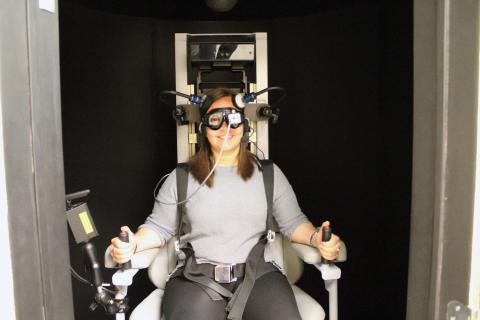
Rotary chair testing is also used to assess the functioning of the inner ear. It is different than VNG because instead of using temperature changes to stimulate the inner ear, whole body rotations are used instead. This is done in a mechanical chair that is situated within a dark chamber. Eye movements are again recorded with infrared goggles. When the chair is rotated, the inner ear senses the rotation, and moves the eyes in the opposite direction. Several different types of rotations are used as a stimulus. In the first, sinusoidal (back and forth) movements of the chair are used at different frequencies (similar to speed, this is the number of rotations per second). A second stimulus that is used is called step velocity testing, or impulse testing. In this part, the chair is rotated at a set velocity for enough time for the nystagmus to taper off (usually 1-2 minutes). By measuring certain variables, such as gain (ratio of eye movements to head movements), phase (how long it takes the eyes to move relative to the head movement), asymmetry (the difference in response between the two ears), and time constant (how long it takes the nystagmus speed to decay to 37% of its original value), these tests can help us determine if the inner ears are functioning properly, or if one or both of the inner ears are weak. In additional, optokinetic nystagmus is tested as well, which measures how the eyes move in response to a moving visual field. This test can be abnormal is certain central conditions, and with bilateral vestibular loss.
Click here to view a video about Rotary Chair Testing.
Vestibular Evoked Myogenic Potentials (VEMP)
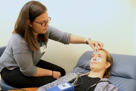
Vestibular Evoked Myogenic Potentials (VEMP) testing is used to assess the otolith organs: the utricle and saccule. Both of these organs are located in a part of the inner ear called the vestibule. The utricle is oriented in the horizontal plane, and senses horizontal linear acceleration (like accelerating in a car), and head tilt. The saccule is oriented in the vertical plane, and therefore senses vertical acceleration (like going up and down in an elevator). Because these organs may have originally been used for sound or vibration detection, they do have some sound sensitivity, which can be measured. Therefore, the test involves placing sound in the ears, and then measuring certain muscle responses to that sound. In a cVEMP (cervical VEMP), sound is delivered to the ear and activates the saccule, which then inhibits some muscles in the neck, including the SCM (sternocleidomastoid) muscle. Because the signal that gets sent to the muscle is inhibitory, the test is done with the muscle contracted, which can be achieved by having the patient lift their head up off the bed during the testing. Normally, the evoked response (electrical change in muscle activity) can only be seen with pretty loud sounds (stimulation levels above 80 dB), but with superior canal dehiscence syndrome, responses are seen at lower levels (60-70 dB). The test can fail to show a response in a variety of conditions, including conductive hearing loss, poor muscle tone, and conditions that damage the saccule like Menière’s disease. Another type of VEMP is the oVEMP (ocular VEMP). This tests the utricle, and sound delivered to the utricle results in excitation of the contralateral eye muscles. Therefore, electrodes placed under the eye, while the patient looks up, are used to measure evoked potentials in the inferior oblique muscle (an eye muscle close to the skin surface). Loss of responses may indicate conductive hearing loss or utricular damage, whereas responses with abnormally high amplitudes (the degree of response) can indicate 3rd window physiology, as occurs with superior canal dehiscence syndrome.
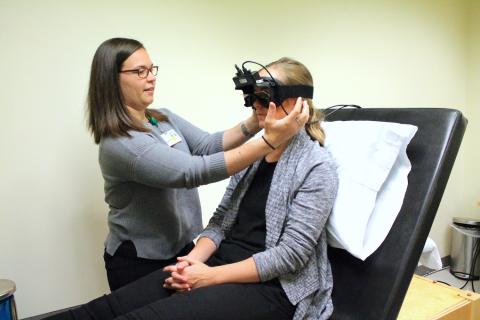
Video Head Impluse Testing (vHIT)
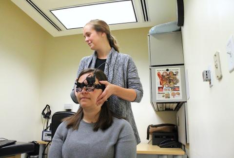
Head impulse testing is used to measure the responses of the three semicircular canals to quick head rotations. The semicircular canals are the part of the inner ear that sense head rotation, and since the inner ears wants to be able to measure rotation in any direction, we have 3 canals in each ears, oriented at 90° to each other. Quick head movements (also called impulses), result in eye movements in the opposite direction. By using a video camera and computer software, we are able to measure quick eye movements that are difficult to see with the naked eye. By rotating the head quickly in the same plane as the semicircular canal being tested, we are able to separately assess each of the six semicircular canals. This test is important for determining if there has been any damage to each of the six semicircular canals, and also for assessing the central compensation that occurs after damage has been done.