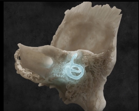
Superior canal dehiscence syndrome (SCDS) was first described by Dr. Lloyd Minor in 1998. Normally, the entire inner ear (a fluid filled space deep within the temporal bone) is encapsulated in dense bone called the otic capsule. In two areas, the oval window (where the stapes sits), and the round window, the inner ear is covered with membranes instead of bone. This design allows for the transmission of sound energy from the ear bones (ossicles) to the inner ear via the oval window, with the round window acting as a low resistance outlet to permit sound energy to vibrate the fluids and membranes of the hearing part of the inner ear- the cochlea- which is a spiral offshoot located between the oval and round windows. Because of this anatomy, the balance part of the inner ear, which is also contiguous with the cochlea, does not experience any fluid vibrations, because it’s not located along this path of least resistance between the oval and round windows. In SCDS, for unknown reasons, bone is absent over the superior semicircular canal (one of the balance organs that senses head rotations in the vertical plane). Since this opening (dehiscence) is the third opening of the otic capsule bone (after the oval and round windows), it’s called a 3rd window. Functionally, this results in two problems. Firstly, pressure transmissions, from either sound or pressure changes (like coughing, straining, or sneezing) can travel between the oval window and the dehiscence, activating the superior canal along the way, resulting in the sensation of movement (vertigo) with sound or pressure changes. Secondly, the 3rd mobile window changes the impedance of the inner ear, making it easier for sounds coming from the body to be heard. This results in several curious symptoms, such as autophony (hearing one’s voice abnormally loudly), pulsatile tinnitus (hearing your heartbeat or pulse in your ear), and hearing other bodily sounds (such as your eyes moving or your neck muscles creaking). Testing for SCDS involves imaging (CT scan), VEMP testing, audiometry (hearing test), and a physical exam. Treatment ![]()
![]() involves surgery, with plugging and/or resurfacing of the dehiscent (open) canal.
involves surgery, with plugging and/or resurfacing of the dehiscent (open) canal.
