
3D Models in Neurosurgery
The Virtual Reality Anatomic Lab at UCSF's Department of Otolaryngology - Head and Neck Surgery offers an innovative approach to understanding complex neuroanatomy. This cutting-edge resource provides detailed 3D models of various anatomical structures, allowing medical professionals and students to explore the intricacies of the human body in unprecedented detail. Dr. Roberto Rodriguez Rubio, an assistant professor in the Department of Neurosurgery, has been pioneering the use of structured light scanning and photogrammetry to revolutionize the depiction of neuroanatomy since 2017.
The lab's extensive collection covers a wide range of anatomical regions, including the brain, skull, head, neck, and spine. Each section features multiple detailed models that showcase different aspects of the anatomy. For example, the brain models include visualizations of the cerebral arteries, venous structures, white matter tracts, and specific regions like the brainstem and cerebellum. Similarly, the skull and head models offer in-depth views of bones, muscles, and neurovascular structures, providing a comprehensive understanding of these complex areas.
This virtual reality approach to anatomical study represents a significant advancement in medical education and surgical planning. By offering highly detailed, interactive 3D models, the Virtual Reality Anatomic Lab enables healthcare professionals to gain a more thorough understanding of human anatomy, potentially leading to improved patient care and surgical outcomes. This resource demonstrates UCSF's commitment to leveraging cutting-edge technology in medical education and practice.
Resident Corner: Endonasal Approach Specific Anatomy
Director and Co-Directors
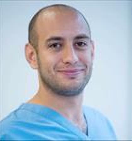
Roberto Rodriguez Rubio, MD

Ivan El-Sayed, MD
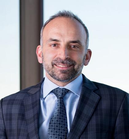
Philip Theodosopoulos, MD

Mitchel Berger, MD
Faculty

Jose Gurrola II, MD

Patricia Loftus, MD

Manish Aghi, MD, PhD, MAS
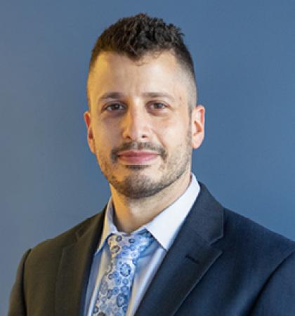
Ezequiel Goldschmidt, MD, PhD
Chairs

Edward Chang, MD
