We provide our patients with multidisciplinary care programs that allow face-to-face collaboration between cancer care specialists. Our Head and Neck surgeons work closely with our colleagues from Head and Neck Reconstructive Surgery, Radiation Oncology, Medical Oncology, Pathology, and Radiology to form specialized Tumor Boards dedicated to delivering the highest quality of cancer care. We are dedicated to the treatment of our patients, partnering with renowned physicians on multidisciplinary tumor boards and providing our areas of expertise in the field of head and neck oncology.
UCSF Head and Neck Oncology Multidisciplinary Tumor Boards
- Head and Neck Tumor Board
- Thyroid Tumor Board
- Visible Tumor Board
- Melanoma Tumor Board
We participate in a number of Institutional and National Clinical Trials aimed to advance cancer care. For more information, please visit our Clinical Trials section.
Areas of Expertise
- The most common type of cancer is squamous cell carcinoma, which originates from the mucosa of the oral cavity.
- Mouth cancer is most often associated with a history of smoking cigarettes/cigars, drinking alcohol, or chewing tobacco.
- Common symptoms include oral pain, bleeding, ulceration, difficulty opening your mouth or moving your tongue, tooth pain, foul breath, and difficulty eating or speaking. If you note any of these symptoms, speak to your primary care doctor or dentist.
- Treatment of cancers of the mouth/oral cavity almost always includes surgery, possibly followed by radiation and/or chemotherapy.
- Good dental hygiene is paramount. If you have teeth in stages of decay, we will have you see our dental department before treatment for evaluation and management.
- Following surgical removal of your tumor, a variety of reconstruction options exist. Your surgeon will talk to you about the best plan for you is in order to restore form and function.
- Maintaining, to the best of our ability, your swallow and speech function is of our utmost importance. We will have you see a member of our Speech Language Pathology team before, during, and after treatment to maximize your function.
The oropharynx is located in the back of the mouth. Cancers in the oropharynx generally arise from lymphoid tissue in the tonsils, adenoids (back of the nose), or base of the tongue.
- The most common type of cancer is squamous cell carcinoma, which originates from the mucosa of the tonsils or base of tongue.
- There has been a rising incidence of oropharynx cancer due to infection with cancerous subtypes of the sexually transmitted Human Papilloma Virus (HPV) in the lymphoid tissue of the tonsils and base of tongue. Your surgeon may ask you about your sexual history, and discuss the need to test a piece of your tumor to confirm HPV infection.
- Smoking and/or drinking associated tumors are less common in the oropharynx, but when cancer is present, these habits may make the tumor more aggressive.
- Many patients with cancers of the oropharynx first notice a neck mass before developing symptoms in their mouth. Any new lump or bump in your neck should be evaluated by a physician.
- Depending on the size and location of the primary tumor, as well as degree of cancer spread to the neck lymph nodes, treatment can consist of a combination of surgery, radiation, or chemotherapy. Discussion of your case at a multidisciplinary tumor conference will help plan your personalized care.
- Maintaining, to the best of our ability, your swallow and speech function is of our utmost importance. We will have you see a member of our Speech Language Pathology team before, during, and after treatment to maximize your function.
Cancer of the head and neck generally arises from mucosal tissue of the mouth and throat. Cancer of unknown primary occurs when cancer is identified in a neck mass, but it is unclear where in the mouth/throat the cancer originated.
- Cancer of unknown primary is most commonly squamous cell carcinoma.
- The surgeon’s goal is to identify the site of origin (also known as the “primary” site). Your surgeon may use a variety of procedures and tests to help with this, including in-office endoscopy and radiology (CT, MRI, PET/CT).
- When our clinic exam and radiology imaging are not able to identify the site of origin, further procedures may be warranted. As most cancers of unknown primary begin in the tonsils or base of the tongue, your surgeon may recommend surgical removal of the tonsils and mucosa over top the base of tongue to help find the site of origin.
- Treatment of cancer of unknown primary is highly patient specific. Individualized treatment plans will be made after discussion of your case and all relevant information (clinic exams, radiology studies, pathology) at a comprehensive multidisciplinary tumor conference.
- Maintaining, to the best of our ability, your swallow and speech function is of our utmost importance. We will have you see a member of our Speech Language Pathology team before, during, and after treatment to maximize your function.
Historically, tumors of the back of the mouth, base of tongue, and throat were removed via large neck incisions, or through the mouth only after cutting the jaw to gain access. Now, with advances in surgical equipment, we are able to safely and effectively remove these tumors through the mouth with a state-of-the-art surgical robot. With the DaVinci Robot, a camera with 3D capability is passed into the mouth to visualize the tumor. From the surgical console, the surgeon is able to manipulate two robotic surgical arms to help in removal of tumors in small, difficult to access locations of the mouth and throat.
- Candidates for TORS often have early stage, small tumors of the tonsils, base of tongue, or throat without spread to neck lymph nodes. Your surgeon will review your case and determine if the robot may be useful.
- TORS offers the advantage of avoiding external neck and/or jaw incisions, and minimizing post-operative speech and swallow functional deficits.
- Maintaining, to the best of our ability, your swallow and speech function is of our utmost importance. We will have you see a member of our Speech Language Pathology team before, during, and after treatment to maximize your function.
Larynx cancer comprises all tumors that arise from the supraglottis (above your voice box), glottis (voice box), subglottis (below your voice box), and hypopharynx (surrounding your voice box, near the entrance to your esophagus).
- The most common type of cancer is squamous cell carcinoma, which originates from the mucosa of the larynx.
- Cancer of the throat is highly associated with a history of smoking cigarettes and drinking alcohol.
- Usually the first symptom is hoarseness, and may be diagnosed as “laryngitis”. Any throat issue that persists after 3 weeks should be evaluated further.
- Other symptoms include difficultly breathing, strained breathing, pain with swallowing, sensation of food/liquid getting stuck on swallowing, throat pain, and voice change. If you note any of these symptoms, speak to your primary care doctor.
- Treatment may consist of a combination of surgery, radiation, and/or chemotherapy. In the event that surgery involves removal of your voice box, our surgeons and voice rehabilitation practitioners are experts in methods to restore intelligible speech.
- Maintaining, to the best of our ability, your swallow and speech function is of our utmost importance. We will have you see a member of our Speech Language Pathology team before, during, and after treatment to maximize your function.
There are four major salivary glands of the head and neck responsible for the vast majority of saliva output to the mouth. These are the paired parotid glands (located in your cheeks in front of your ears) and paired submandibular glands (located underneath your jaw bone on either side of your neck). In addition, there are hundreds of small mucous producing glands lining the mucosa of your lips, tongue, palate, and inner cheeks. Both benign and malignant tumors affect the salivary glands.
- Fortunately, most tumors of the major and minor salivary glands are benign. These include pleomorphic adenomas, Warthin tumors, basaloid adenomas, and oncocytomas, among others.
- Cancerous tumors more often affect the minor salivary glands, but can also be found in the major salivary glands. These include mucoepidermoid carcinoma, adenoid cystic carcinoma, and acinic cell carcinoma, among others.
- Other than smoking (which is associated with benign Warthin tumors), there are no other known environmental risk factors that may cause salivary gland cancer.
- Tumors of the salivary glands are most often noticed by the patient as a new lump or bump in front of the ear, or near the angle of you jaw. Symptoms that are concerning and may suggest cancer include pain, numbness of the face, or weakness of facial muscles.
- A fine needle aspiration biopsy is often ordered to help diagnosis the type of tumor and determine if it is cancerous.
- Treatment of benign and malignant salivary gland tumors often involves surgery.
As one of the first centers in the United States to pioneer sialendoscopy techniques, the UCSF Sialendoscopy Center is globally recognized as a leader in the development and advancement of this minimally invasive therapy. Prior to sialendoscopy, treatment for salivary gland obstruction due to stones was limited to surgical removal of the affected gland. Modern technology allowed for the development of extremely small endoscopes (size range: 0.8mm to 1.6mm). Our experts use this small endoscopic camera system to enter the salivary ducts through the mouth and examine the duct system for problems such as salivary duct stones (sialolithiasis) and scarring (stenosis). Our providers are able to use the sialendoscope to guide forceps and wire baskets to remove salivary stones and break up larger stones using a laser (lithotripsy). Sialendoscopy also allows for the application of medications for treatment of blocked salivary ducts. Often these treatments involve limited or no incisions in the mouth and can be an alternative to open surgery or salivary gland removal.
Our sialendoscopy experts train head and neck surgeons around the nation on cutting-edge sialendoscopic techniques and are actively engaged in advancing the field through innovation and research. Some of their studies have addressed:
- The combination of sialendoscopy with ultrasound imaging for diagnosis and therapies.
- Predicting which patients would benefit from sialendoscopic surgery.
- Following patients with stones and stenosis to study the benefits of sialendoscopy.
- Surgical techniques for accessing the salivary ducts.
- Pioneering new techniques to remove and break apart salivary stones.
Skin cancer is now the most common diagnosed cancer in the US. Each year there are more new cases of skin cancer than new cases of breast, prostate, lung, and colon cancer combined.
- The most common types of skin cancer are melanoma, basal cell carcinoma, and squamous cell carcinoma.
- The number one risk factor for all forms of skin cancer is ultraviolet radiation, which is found in natural sun light and tanning beds.
- While melanoma accounts for a very small percentage of all skin cancers, it is potentially the most dangerous. They often first appear as small moles or brown spots. It is important to know warning signs that may help you get treated early. A helpful mnemonic is “ABCDE” – “A”: asymmetry of mole (it is not perfectly round); “B”: borders are not smooth and even; “C”: the color is not uniform, or it is darker; “D”: the diameter is larger than a pencil tip; “E”: the mole is evolving, or changing over time. Contact your doctor if you notice any of these characteristics in a mole.
- Skin cancer prevention is paramount. Always where sunscreen, avoid prolonged direct sunlight to your skin, and do not use tanning beds.
Minimally invasive skull base surgery (MISB) is a rapidly evolving field applying endoscopic technology to remove tumors in the head and neck, paranasal sinuses, skull base, and intracranial compartment. The technique allows our surgeons to more easily access both benign and malignant tumors in previously inaccessible locations.
- Both benign and malignant tumors may arise in the skull base and paranasal sinuses. Common benign tumors include juvenile nasopharyngeal angiofibromas, meningiomas, chordomas, and schwannomas, as well as tumors arising from the pituitary gland. Common malignant tumors include metastases from other sites of the body, melanoma, squamous cell carcinoma, and ethesioneuroblastoma.
- MISB surgery uses an endoscope – a long, flexible instrument attached to a camera and a light source – to guide surgeons through very small ports of entry to remove these tumors.
- These procedures involve very small or no external incisions, resulting in fewer complications, reduced blood loss, and shorter recovery times for patients.
- The most common approach is through the nose and involves the combined efforts of an Otolaryngologist and a Neurosurgeon in the operating room.
Thyroid and parathyroid surgery consists of complex, highly-skilled surgery to treat a broad spectrum of benign and malignant conditions of the thyroid & parathyroid glands. From simple removal of suspicious thyroid nodules or parathyroid glands, to removal of massive multinodular goiters, to the most advanced thyroid cancer surgery possible, we specialize in comprehensive, high-volume thyroid & parathyroid surgery.
- Thyroid Cancer: Our team’s thyroid/parathyroid surgeons are all members of the UCSF Helen Diller Family Comprehensive Cancer Center. We take a personalized, careful approach to all patient care, and discuss our cases in multidisciplinary thyroid cancer conference. While simple removal of a thyroid lobe is our most common surgery, we specialize in advance thyroid cancer surgery where the disease is high-risk and the need for cure and prevention of recurrence is of critical importance. From comprehensive removal of regionally metastatic lymph nodes in the lateral neck, to central neck surgery for recurrent/persistent thyroid cancer, to surgical and non-surgical treatment of widely invasive thyroid cancers, we manage thyroid cancer with the highest level of skill and precision in the world.
- Thyroid Nodules and Goiters: We offer surgery for simple thyroid nodules in order to achieve diagnosis that cannot be made through fine needle aspiration (FNA) biopsy. We also offer therapeutic surgery for massive multinodular goiters compressing the neck, airway, and other structures.
- Parathyroid Disease: We offer comprehensive management of all parathyroid disease, including surgery for primary, secondary, and tertiary hyperparathyroidism. We use a minimally-invasive approach and intraoperative PTH monitoring to provide biochemical evidence of disease cure by the time our patients reach the recovery room.
- Ultrasound and fine needle aspiration biopsy: Our surgeons offer in-clinic ultrasound of the thyroid and parathyroid glands and neck lymph nodes with real-time characterization of the patient’s relevant anatomy, concerning thyroid nodules & lymph nodes. We use ultrasound findings to personalize treatment plans for each patient in the first clinic visit. With the help of on-site UCSF cytopathology experts skilled in thyroid & parathyroid pathologic diagnosis, we offer bedside needle biopsy during the clinic visit. Combined with in-clinic ultrasound, these can help the surgeon and patient create an accurate, tailored plan of care for each patient.
We take great pride in providing outstanding thyroid/parathyroid surgical care. Our primary goal is to work tirelessly to afford each patient the highest chance of cure with the lowest risk of complications or disease recurrence.
Microvascular head and neck reconstruction is a technique for rebuilding the face and neck using blood vessels, bone, and tissue from other parts of the body. The technique is one of the most advanced surgical options available for rehabilitating surgical defects that are caused by the removal of head and neck tumors.
- The technique involves harvesting flaps of healthy tissue from remote parts of the body – where the tissue is not that important – and then transferring them to the wound bed. They are also known as “free flaps”, as the tissue is taken from one part of the body and transferred to a new part that is in need of reconstruction.
- Common areas of the body to acquire free flaps include the lower leg, lateral thigh, forearm, and back.
- A microscope is used to suture the blood vessels of the flap to blood vessels in the neck, allowing the tissue to live as if it were back in its original location.
- Donor sites of for the free flap are chosen with respect to each patient to both optimize the reconstruction and limit the donor site morbidity
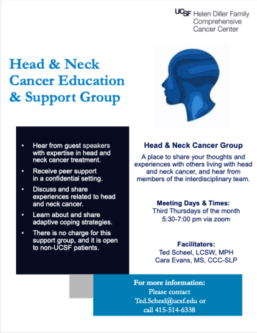
Head & Neck Cancer Support Group

